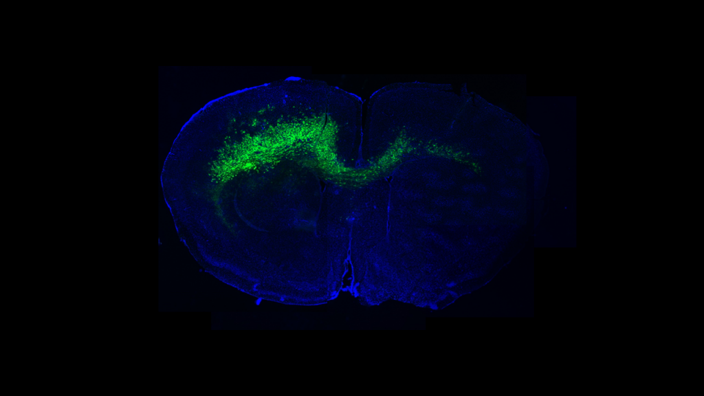
Glioblastomas are among the most deadly cancers, full stop. With an average five-year survival rate of just 5% to 7%, research and innovation in the treatment of these tumors is essential. There has been rapidly growing interest in a critical interplay between glioblastomas and the surrounding neurons: neuronal activity has been established as a driver of glioma proliferation and infiltration [1], leading these tumors to progress faster and become much harder for a surgeon to resect. Glioblastomas are also known to drive hyperexcitability through the release of toxic levels of glutamate [2], setting the stage for a pernicious positive feedback loop. It is for these reasons that a compound capable of putting the brakes on this process is of utmost interest in the treatment of gliomas.
To address this, I generated a peptide (affectionately nicknamed “Kegstand”) against Note: the glypican 5 used as bait was generated in eukaryotic cells (mouse) as we suspect the glycosylation may play a role in its effect on synapses.. The reasons glypican 5 makes a good target, I reasoned, are:
- It is emerging that glypican 5 plays a role in the stabilization and strength of synaptic connections [3].
- Glypican 5 appears to internalize slowly and only to a limited extent relative to classic internalizing drug targets, allowing me to sidestep the infamous “binding-site barrier” that plagues targeted drug delivery [6], [7]. Because my payload is designed to act through a bystander effect rather than requiring receptor-mediated uptake, this limited internalization is a feature rather than a liability.
- Several glypican-family proteins are implicated in stress-response pathways [8], [9], and evidence from past projects suggests that glypican-5 may be upregulated in regions of high cellular stress (neurosphere and colocalization data), implying that glypican 5 is likely abundant in these hyper-excitable, glutamate-rich milieux [4], [10].
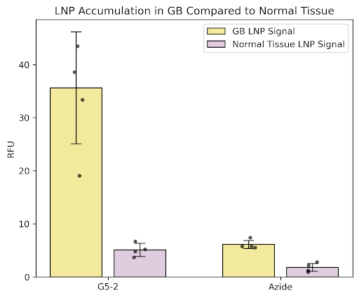
It is important to keep in mind that these errant, over-excited neurons in the tumor margins are not themselves unique, and thus any activity my peptide has on them would also translate to normal, healthy neurons — not ideal. This is why I use a multimerization scheme that builds off of the work of Kathlynn Brown [5] to keep distribution of this peptide local to the tumor and away from healthy tissue.
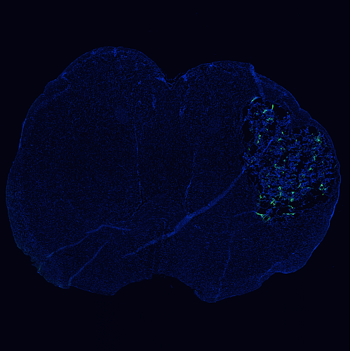
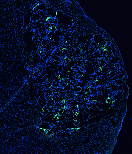
As part of a “reverse rescue”–style experiment, I divided otherwise identical plates of neurons into three groups: one that received additional soluble glypican 5 on the first day in vitro (DIV), one that received identical glypican 5 plus a daily administration of Kegstand peptide, and one cultured according to standard neuron culture protocol as a control. I then fixed and stained for synapses and nuclei; the images were then quantified by number of synapses per neuron to investigate both:
- the connection between glypican 5 and synaptic formation, and
- the effect my peptide, Kegstand, has on neuronal processes and structures.
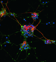
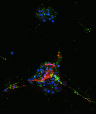
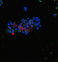
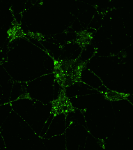
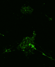
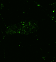
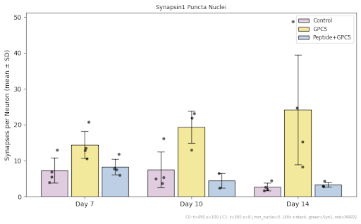
The data shows that neurons cultured in the presence of extra glypican 5 do indeed form synapses more readily than the control neurons at the same time points, with that difference only increasing at later time points. The neurons that received both excess glypican 5 and Kegstand peptide showed only slightly elevated synapse formation relative to the control group.
Investigation into Kegstand’s effects on the electrical activity of neurons is currently ongoing as of August 2026; beyond this, I have studies planned to probe these questions in vivo.
More to come…
References
- Venkatesh HS, Morishita W, Geraghty AC, et al. Electrical and synaptic integration of glioma into neural circuits. Nature. 2019;573(7775):539–545. https://doi.org/10.1038/s41586-019-1563-y ↩
- Buckingham SC, Campbell SL, Haas BR, Montana V, Robel S, Ogunrinu T, Sontheimer H. Glutamate release by primary brain tumors induces epileptic activity. Nat Med. 2011;17(10):1269–1274. https://doi.org/10.1038/nm.2453 ↩
- Bosworth AP, Contreras M, Sancho L, Salas IH, Paumier A, Novak SW, Manor U, Allen NJ. Astrocyte glypican 5 regulates synapse maturation and stabilization. Cell Rep. 2025;44(3):115374. https://doi.org/10.1016/j.celrep.2025.115374 ↩
- Yu K, Lin CCJ, Hatcher A, Lozzi B, Kong K, Huang-Hobbs E, et al. PIK3CA variants selectively initiate brain hyperactivity during gliomagenesis. Nature. 2020;578(7793):166–171. https://doi.org/10.1038/s41586-020-1952-2 ↩
- Gray BP, Li S, Brown KC. From phage display to nanoparticle delivery: functionalizing liposomes with multivalent peptides improves targeting to a cancer biomarker. Bioconjug Chem. 2013;24(1):85–96. https://doi.org/10.1021/bc300498d ↩
- Fujimori K, Covell DG, Fletcher JE, Weinstein JN. A modeling analysis of monoclonal antibody percolation through tumors: a binding-site barrier. J Nucl Med. 1990;31(7):1191–1198. https://jnm.snmjournals.org/content/31/7/1191 ↩
- Thurber GM, Schmidt MM, Wittrup KD. Antibody tumor penetration: transport opposed by systemic and antigen-mediated clearance. Adv Drug Deliv Rev. 2008;60(12):1421–1434. https://doi.org/10.1016/j.addr.2008.04.012 ↩
- Ebong EE, Lopez-Quintero SV, Rizzo V, Spray DC, Tarbell JM. Shear-induced endothelial NOS activation and remodeling via heparan sulfate, glypican-1, and syndecan-1. Integr Biol (Camb). 2014;6(3):338–347. https://doi.org/10.1039/c3ib40199e ↩
- Cheng F, Mani K, van den Born J, Ding K, Belting M, Fransson LÅ. Nitric oxide-dependent processing of heparan sulfate in recycling S-nitrosylated glypican-1 takes place in caveolin-1-containing endosomes. J Biol Chem. 2002;277(46):44431–44439. https://doi.org/10.1074/jbc.M205241200 ↩
- Chung WJ, Lyons SA, Nelson GM, Hamza H, Gladson CL, Gillespie GY, Sontheimer H. Inhibition of cystine uptake disrupts the growth of primary brain tumors. J Neurosci. 2005;25(31):7101–7110. ↩
- Huang-Hobbs E, Cheng YT, Ko Y, Luna-Figueroa E, Lozzi B, Taylor KR, McDonald M, He P, Chen HC, Yang Y, Maleki E, Lee ZF, Murali S, Williamson MR, Choi D, Curry R, Bayley J, Woo J, Jalali A, Monje M, Noebels JL, Harmanci AS, Rao G, Deneen B. Remote neuronal activity drives glioma progression through SEMA4F. Nature. 2023;619(7971):844–850. https://doi.org/10.1038/s41586-023-06267-2 ↩